
Fig. 2.
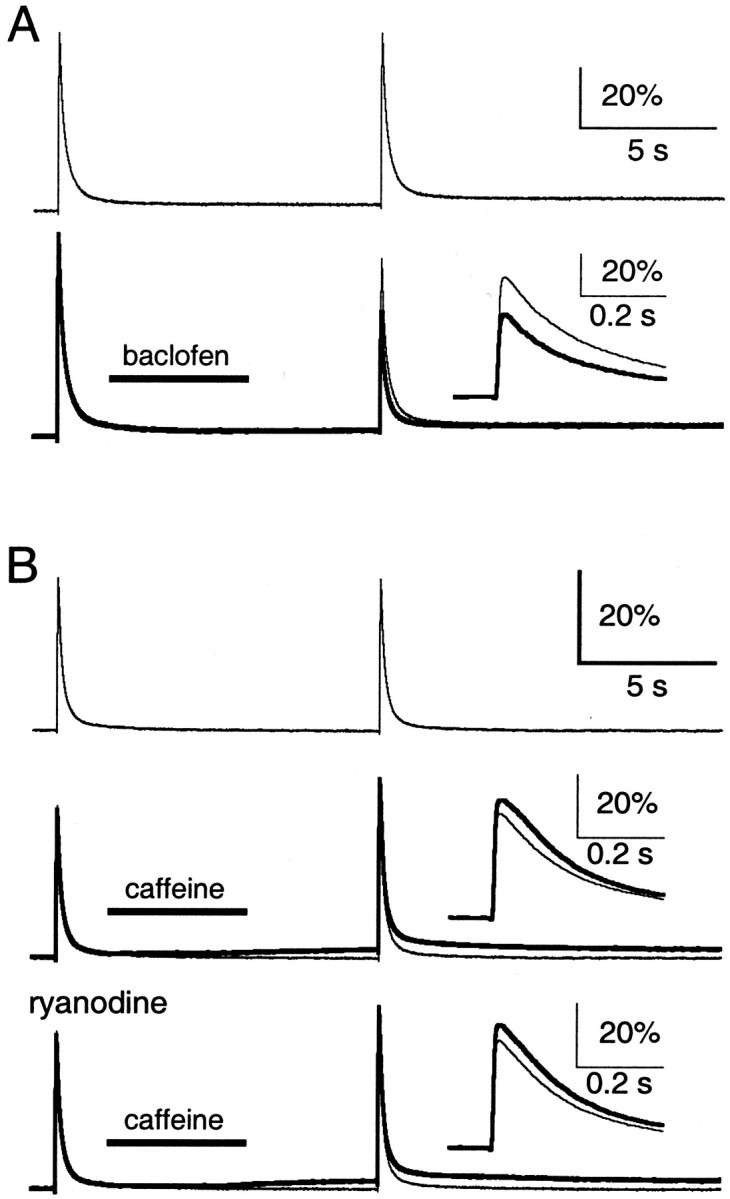
Lack of caffeine-evoked CICR at the parallel fibers. Parallel fibers were filled with Oregon Green 488 BAPTA-1 AM. In each trial, parallel fibers were stimulated with a control and test stimulus separated by 10 sec. A, ΔF/F signals in the absence of drug application (top, bottom, light traces) and with puff application of 500 μm baclofen (bottom, bold trace).Inset, Test ΔF/F signals with baclofen (bold trace) or without (light trace) on an expanded time scale. B, ΔF/F signals in the absence of drug application (top, middle, bottom, light trace) and with puff application of 40 mm caffeine, in the absence (middle, bold trace) and presence (bottom, bold trace) of 10 μm ryanodine. Insets, Test ΔF/F signals with caffeine (bold trace) or without (light trace) on an expanded time scale. Representative traces are averages of three to five trials. For insets in B, the slow ΔF/F signal has been subtracted.