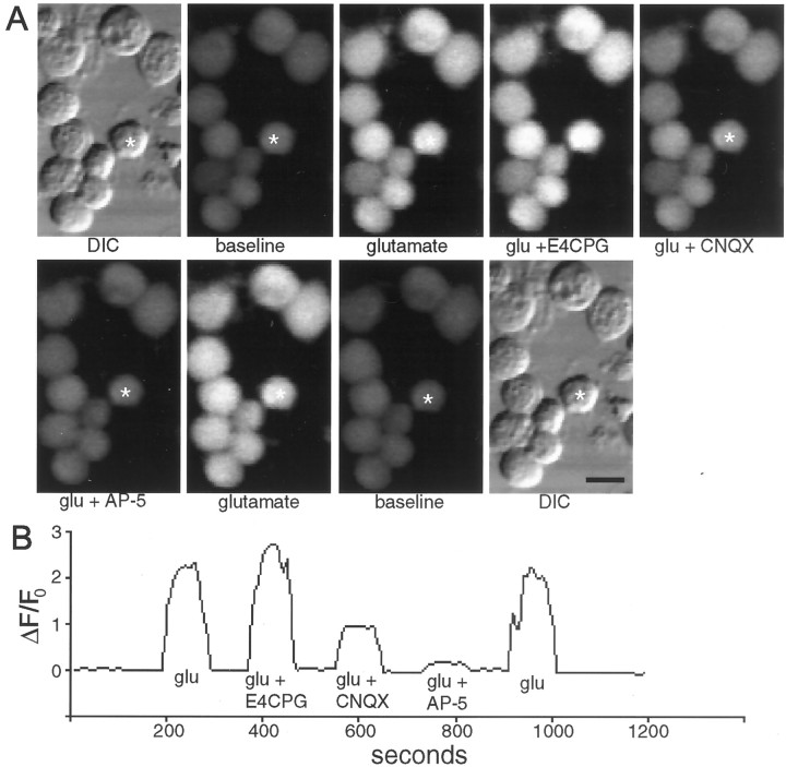
Fig. 2.
Fluo-3 AM imaging of the calcium response to glutamate and its reduction by glutamate receptor antagonists. A, Representative DIC and fluorescent images of Xenopus tectal neurons under various stimulation conditions. CNQX and AP-5 reduce the fluorescence increase in response to glutamate, whereas E4CPG increases the calcium response without permanently altering the baseline fluorescence or impairing the ability of the cell to respond to subsequent applications of glutamate after removal of the antagonists. Scale bar, 10 μm.B, Graph of ΔF/F0 versus time for the single cell marked by the asterisks in Ashowing the quantitation for the glutamate-induced calcium-dependent fluorescence increases in the presence of various glutamate receptor antagonists.

