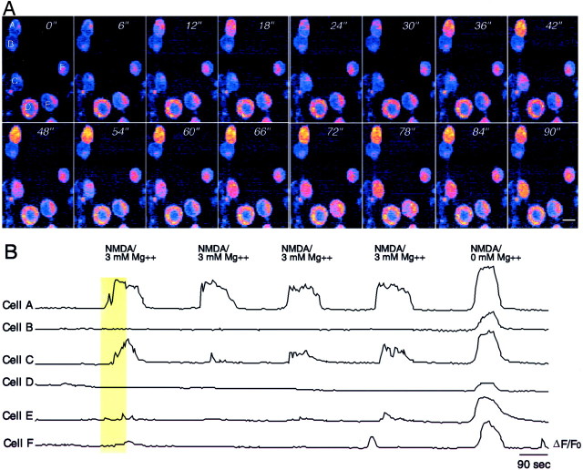
Fig. 6.
NMDARs on some early Xenopus tectal neurons have low magnesium sensitivity. A, Frames showing a field of six neurons at 1 DIV after loading with fluo-3 AM. Images collected at 6 sec intervals illustrate the NMDA-induced calcium fluorescence in a recording solution containing 3 mmmagnesium. Two cells (A, C) have significant calcium influx in response to 100 μm NMDA + glycine in 3 mm magnesium, whereas four other cells show little or no response. B, Plot of ΔF/F0 for the same cells over a longer time interval as well as their response to NMDA in magnesium-free solution. The vertical yellow bar shows the frames of the record illustrated in A. Scale bar, 10 μm.

