Fig. 2.
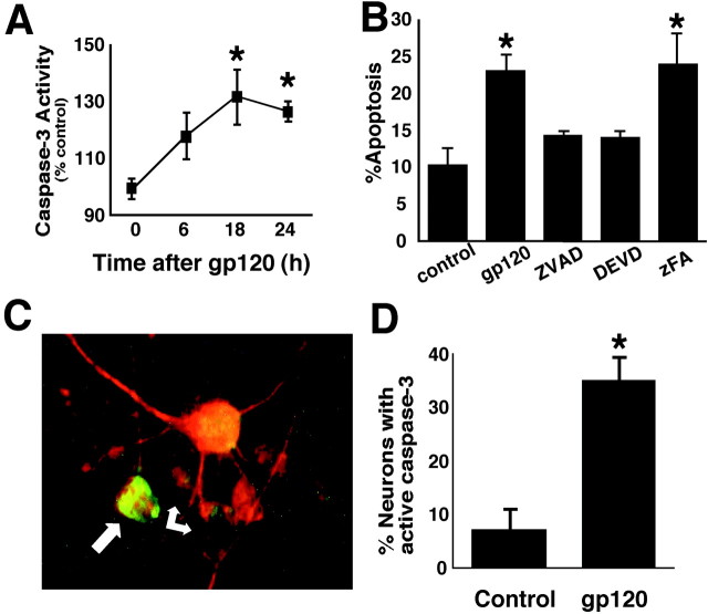
Cultured rat cerebrocortical cells exposed to HIV/gp120 manifest caspase-3 activity and undergo caspase-dependent neuronal apoptosis. A, Caspase-3-like activity (DEVD cleavage) was significantly increased over control cultures after 18–24 hr exposure to 200 pm gp120 (*p< 0.01; n = 3). B, Exposure to gp120 produced a statistically significant increase in apoptosis that was prevented by zVAD-fmk or DEVD-fmk but not by zFA-fmk (n = 9 from 3 independent experiments).C, A number of neurons identified with anti-MAP-2 (red) also contained active caspase-3 (green; arrows). D, The percentage of MAP-2-positive neurons labeled with active caspase-3 greatly increased in cultures exposed to gp120 for 24 hr (*p < 0.001; n = 6 from 2 independent experiments).

