
Fig. 1.
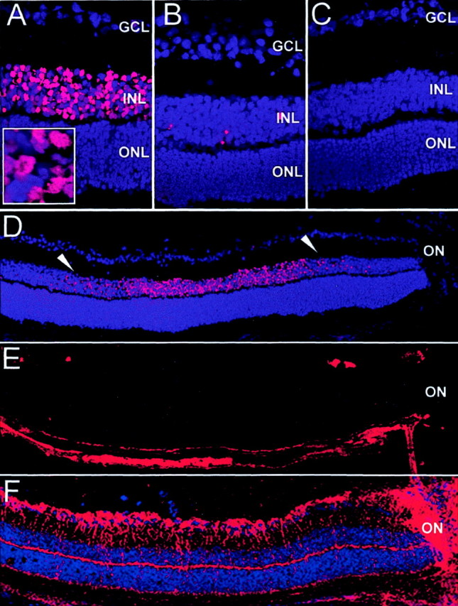
Apoptosis in the central P14 retina detected by TUNEL staining. A–D, TUNEL staining; E, lectin Griffonia simplicifolia; F, GFAP, immunohistochemistry. A, D–F, Eyes of iNOS(+/+) mice of the ischemic phase of the experiment. B, Eyes of iNOS(−/−)mice of the ischemic phase of the experiment. C, Room-air-raised iNOS(+/+) control mice. TUNEL-positive cells were found in the central inner nuclear layer of ischemic iNOS(+/+) sections (A). Room-air-raised iNOS(+/+)mice (C) do not show any TUNEL-positive cells, and ischemic iNOS(−/−) eyes show only very few positive cells (B). The TUNEL-positive cells in the INL of post-oxygen-incubated iNOS(+/+) eyes also exhibited signs of chromatin condensation and pyknotic nuclei (inset in A). The signal on sections of ischemic iNOS(+/+) mice is restricted to the INL of the central retina (D, betweenarrowheads). This area corresponds to the capillary-free retina, as demonstrated by endothelial cell staining (E) in which RMG cell activation can be visualized by GFAP immunohistochemistry (F) (adjacent sections). Experiment represents one of three independent experiments that gave similar results. ON, Optic nerve. Panel height: A–C, 240 μm; inset inA, 20 μm; D–F, 370 μm.