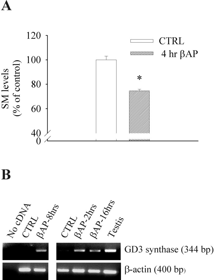
Fig. 3.
A, Exposure of cultured cortical neurons to βAP(25–35) for 4 hr reduces sphingomyelin (SM) levels. Values are means ± SEM of five individual determinations. *p < 0.01 (Student'st test) compared with control cultures.B, Expression of GD3 synthase in primary cultures of rat cortical neurons exposed to βAP(25–35) for the indicated times. The results of two representative experiments are shown.CTRL, Control cultures. Amplification of β-actin cDNA was performed to confirm the integrity of the cDNA preparations and to control for genomic DNA contamination. The 600 bp of β-actin was not detected, thus excluding any genomic contamination.

