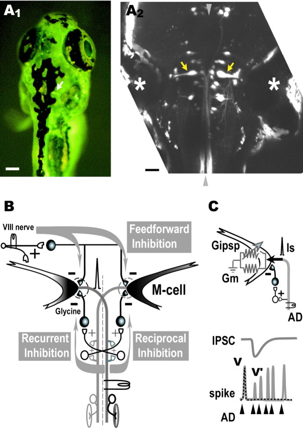
Fig. 1.

Retrograde labeling of the Mauthner cells.A1, Dorsal view of a living zebrafish at 6 dpf that had been injected in the spinal cord with CGD. An M-cell (arrow) can be observed in the intact fish by fluorescence microscopy. Scale bar, 100 μm. A2, Projected confocal stack of the array of reticulospinal neurons in horizontal plane in another larva labeled by bilateral injection of CGD into the spinal cord. The pair of M-cells (arrows) are easily identified as the largest neurons with a lateral dendrite and a commissural axon and are located beside the otic vesicle (asterisk). Rostral is at top.Arrowheads represent the midline. Scale bar, 50 μm.B, The M-cell of an adult zebrafish receives feedback inhibition from the axon collateral as well as feedforward inhibition from eighth nerve afferents. The feedback inhibition consists of recurrent and reciprocal pathways that are mediated by T-reticular neurons (open symbols) and glycinergic interneurons (filled symbols). Note that T-reticular neurons appear to be homologous to cranial relay neurons in the goldfish and giant fiber neurons in the hatchetfish (Kimmel et al., 1985).C, Because the AD action potential of the M-cell propagates almost passively from the initial segment to the soma, the spike amplitudes in the absence (V) or presence (V′) of inhibition are expressed respectively as V =Is/Gm orV′ =Is/(Gm +Gipsp), whereGm and Gipsprepresent the resting and inhibitory synaptic conductances, respectively, and Is represents the spike current. Hence,Gipsp/Gmis estimated from the ratio of V/V′ (cf.Furukawa and Furshpan, 1963; Faber and Korn, 1982; Oda et al., 1995;Hatta and Korn, 1998).
