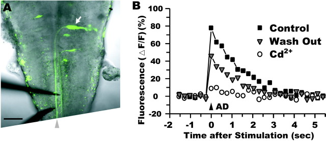
Fig. 3.
The fluorescence change mediated by voltage-activated Ca2+ channels. A, Ventral view of an exposed brain of a zebrafish larva (see Materials and Methods). A fluorescent image of retrograde labeling of a left M-cell (arrow) and a transmitted light image were merged. Bipolar stimulus electrodes (bottom left) were inserted into the caudal hindbrain and positioned near the M-axon (arrowhead). Rostral is at the top. Scale bar, 50 μm. B, The fluorescence increase (square) in response to stimulation of the M-axon was almost completely abolished (circle) within 10 min by bathing with cadmium (30 μm)-containing solution. The response partially recovered (triangle) after 46 min washout. Stimulus intensity was 1.2T in control and washout but 2.5T during cadmium application.

