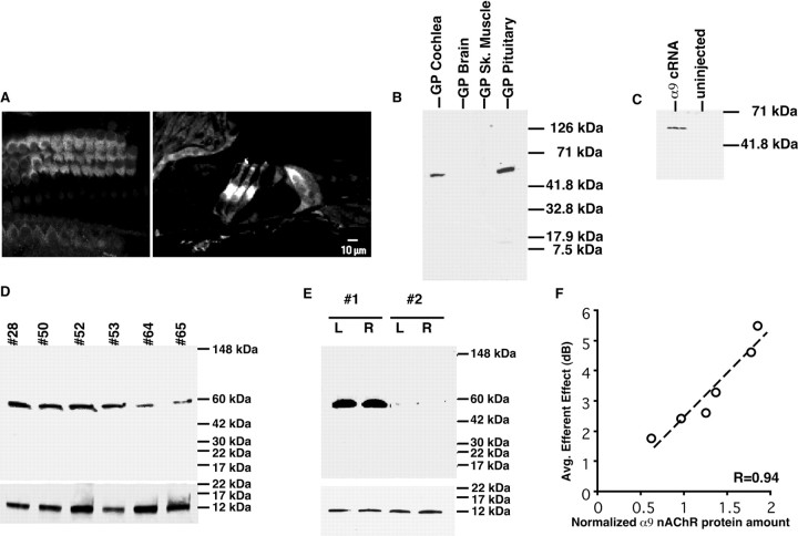
Fig. 5.
A given animal's average efferent strength is proportional to the amount of α9 nAChR present in the cochlea.A, Immunohistochemistry using the α9snAChR antibody (MU43f) on cochlear sections. When the cochlea is imaged in the Z plane, the staining is found in the basal membrane of the outer hair cells. α9s nAChR expression in the basal portion of the three rows of outer hair cells and is present to a lesser extent in inner hair cells. B, α9snAChR is expressed in the cochlea and pituitary gland but not in brain cortex or skeletal muscle tissues by Western blot analysis.C, Western blot using MU43f antibody detects α9s nAChR protein in membranes isolated from α9s-injected but not non-injected control X. laevis oocytes showed that this antibody can recognize heterogeneously expressed α9s nAChR protein.D, Western blot analysis showed the variability of α9s nAChR protein expression in the cochlea of six guinea pigs (40 μl/lane, ∼30 μg of cochlear protein). Bottom blot shows oncomodulin Western blot used to normalize for equal protein loading. E, Western blot analysis showing that, whereas α9s nAChR expression varies between animals (#1, #2), expression was equivalent between the right (R) and left (L) ears of the same animal (n = 4 ears). Bottom blot shows oncomodulin Western blot showing there was no significance difference between oncomodulin protein (and hence cochlear proteins) loaded into each lane of the gel. F, α9s nAChR protein expression correlated with the magnitude of the average efferent strength (r = 0.97; p < 0.002;n = 6 ears). Blots were normalized to the amount of the calcium-binding protein oncomodulin, which is only expressed by outer hair cells (Sakaguchi et al., 1998). The amount of oncomodulin present in each cochlea sample did not vary significantly.

