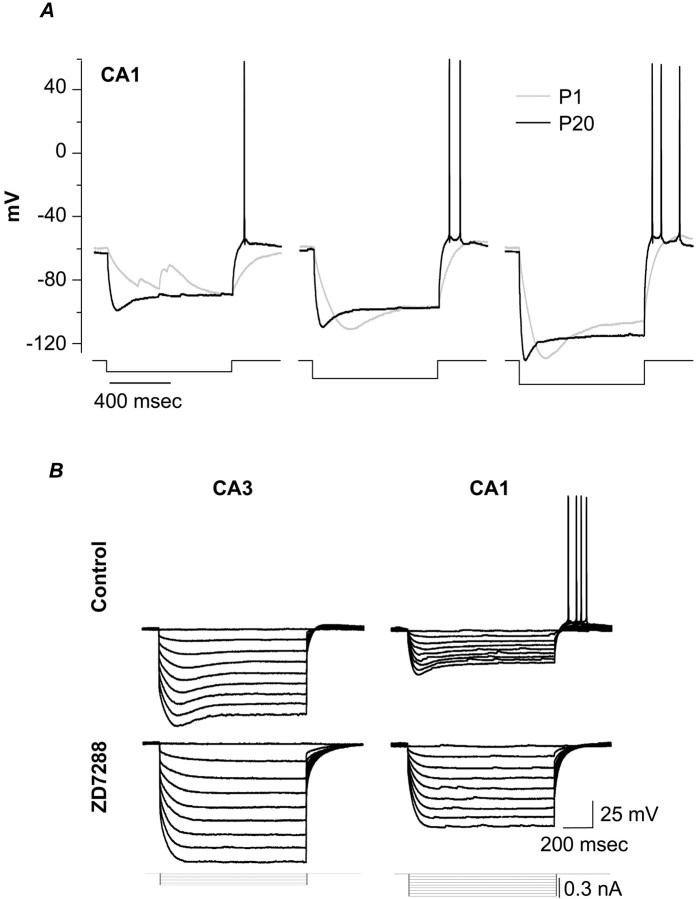
Fig. 1.
A, Developmental changes in responses to hyperpolarizing current injections and in posthyperpolarization rebound firing. These current-clamp recordings show voltage responses in response to 1-sec-long current injections calibrated to yield similar steady-state hyperpolarizations. Current injections are as follows: 10 pA (left), 20 pA (middle), and 30 pA (right) for P1 (gray traces) and 140 pA (left), 200 pA (middle), and 300 pA (right) for P20 (black traces). B, Comparison of voltage sag during hyperpolarizing current injections, and subsequent rebound firing, in representative P20 CA3 (left) and CA1 (right) neurons. Top, Controltraces. Bottom, Tracesrecorded during exposure to the Ih blocker ZD7288 (100 μm, 20 min).

