
Fig. 9.
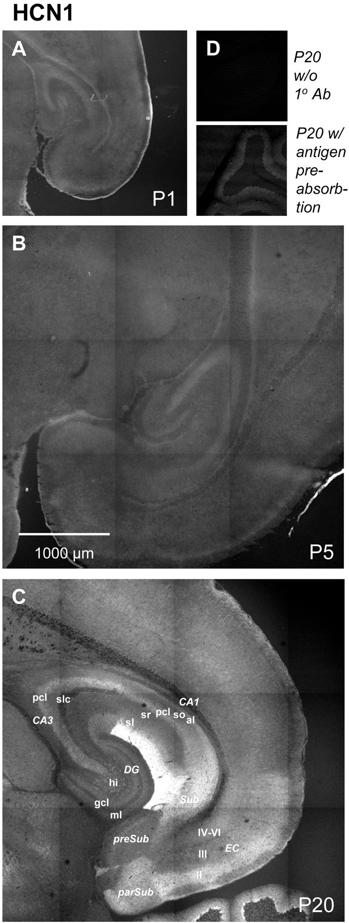
Developmental changes in HCN1 immunoreactivity. Shown are horizontal sections illustrating HCN1 immunoreactivity at P1 (A), P5 (B), and P20 (C). To facilitate qualitative comparisons of immunoreactivity, tissues of different ages were stained simultaneously using an identical protocol, and images were acquired using the same confocal microscope parameters. Negative controls included omission of 1o antibody (Ab) (D,top) and preincubation of antibody with the relevant peptide antigen (D, bottom). Abbreviations are as in Table 2. II, Entorhinal cortex layer II; III, entorhinal cortex layer III;IV-VI, entorhinal cortex layer IV-VI; al, alveus; CA1, CA1 field of the hippocampus;CA3, CA3 field of the hippocampus; DG, dentate gyrus; EC, entorhinal cortex;gcl, granule cell layer; hi, hilus;ml, molecular layer; parSub, parasubiculum; pcl, pyramidal cell layer;preSub, presubiculum; sl, stratum lancosum; slc, stratum lucidum; sm, stratum moleculare; so, stratum oriens;sr, stratum radiatum; Sub, subiculum.