
Fig. 7.
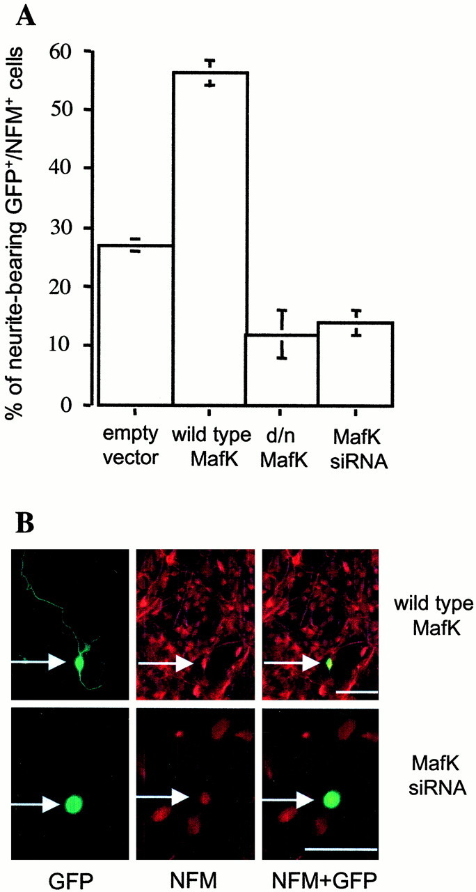
Functional role of MafK in neurite outgrowth by telencephalic neurons. Cells cultured from E14 rat telencephalon were transfected with empty vector, wild-type MafK, d/n MafK, or MafK siRNA plus empty vector. Thirty hours after transfection, cultures were immunostained with antisera against GFP (green) and NFM (red). A, Percentage of neurite-bearing GFP- and NFM-positive cells. Values are averages of two counts representing >50 cells each. Error bars indicate range of counts. Comparable results were achieved in this and an independent experiment by scoring the proportion of neurite-bearing GFP-positive cells. B, Examples of morphology of NFM-positive cells transfected with wild-type MafK and MafK siRNA. Left panels, Staining for GFP; middle panels, staining for NFM; right panels, merged images.Arrows indicate the same cell (left toright) stained with GFP and NFM. Scale bars, 50 μm.