
Fig. 6.
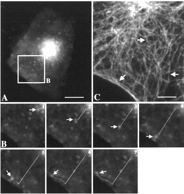
Dynamics of intracellular trafficking of rapsyn–GFP by videomicroscopy: role of microtubules. A, The COS-7 cell transfected with rapsyn–GFP was analyzed by three-dimensional videomicroscopy after 24 hr of expression.B, Time series imaging of the selected area of the cell in A. The organelle indicated by thearrow moved straight from the cell center to the periphery and then tangentially to the cell surface. Both the velocity (∼0.2 μm/sec) of the transport and the linear trajectory suggested a microtubule-guided movement. The trajectory of rapsyn–GFP transporters is consistent with the microtubular organization (C) radiating from the MTOC (arrow) toward the cell periphery and underlying the plasma membrane (arrows). Intervals between frames are 20 sec. Scale bars: A, 10 μm; C, 5 μm.