
Fig. 8.
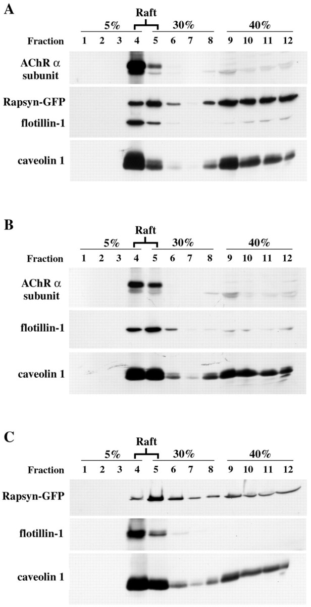
Raft fractionation of rapsyn–GFP and AChR in COS-7 cells. Cells transiently cotransfected with rapsyn–GFP and AChR subunits (A) or separately with AChR subunits (B) or rapsyn (C) were subjected to subcellular fractionation by extraction with 1% (w/v) Triton X-100 on ice. Cell lysates were separated by ultracentrifugation on discontinuous sucrose density gradients. Fractions collected from the top of the gradient were separated by SDS-PAGE (12% acrylamide) and analyzed by immunoblotting (see Materials and Methods). The distributions of rapsyn–GFP and AChR α subunit along the gradient were compared with those of caveolin-1 and flotillin-1, two endogenous markers of lipid rafts. AChR α subunit (49 kDa) and rapsyn–GFP (70 kDa) fractionated mostly in the low-density caveolin-1(26 kDa)/flotillin-1(52 kDa)-enriched raft fractions (lanes 4, 5).