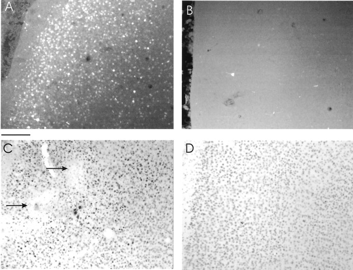
Fig. 1.
Histological examination shows successful expression of transgenes in the primary visual cortex.A, The photomicrograph shows a very large number of cells expressing mCREB–GFP near the site of an injection made 4 d earlier. Scale bar, 180 μm. B, Few cells expressed mCREB–GFP ∼2 mm away from the injection. C, Darkly labeled cells stained with CREB-specific antibodies (1:1000; Upstate Biotechnology) are seen near the injection of HSV–CREB. Note the presence of electrolytic lesions (arrows) made during an electrode penetration near the injection site. D,Lightly stained cells ∼2 mm away from the injection of HSV–CREB. The immunohistochemical procedures were chosen to minimize detection of endogenous CREB and therefore underestimate the number of infected cells.

