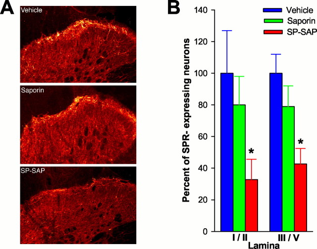
Fig. 1.
Loss of SPR-expressing neurons after intrathecal infusion of SP-SAP. A, Confocal images showing representative examples of SPR-IR in animals pretreated intrathecally with vehicle, SAP alone, or SP-SAP. A dramatic reduction in SPR-IR is evident after SP-SAP. B, Mean ± SEM percentage of neurons that express the SPR after intrathecal vehicle, SAP, or SP-SAP. The number of SPR-expressing cells was obtained from individual animals, and a mean ± SEM was calculated. This mean value was designated as 100%, and the SEM was proportionately adjusted (as a percentage) to provide a measure of variability. Data for SAP- and SP-SAP-treated groups represent the percentage of cells compared with the vehicle-treated group. ∗Significant differences from vehicle.

