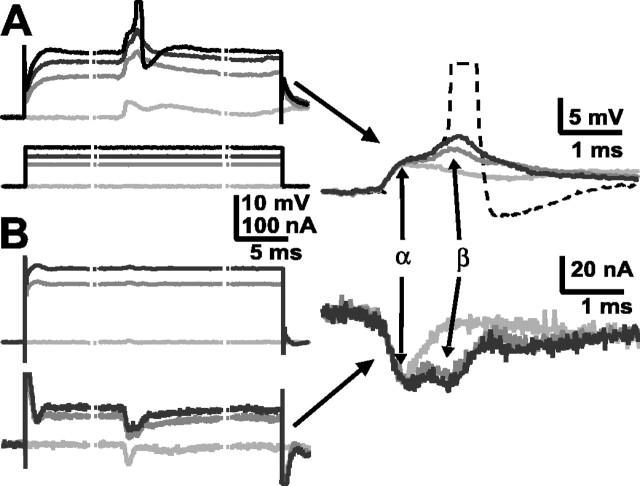
Fig. 7.
Modulation of LG synaptic inputs by LG depolarization. A, EPSPs in LG (top) evoked by nerve stimulation when LG was at rest or depolarized by current injection (bottom). B, EPSCs in the same LG (bottom) evoked by the same nerve stimulation when LG was voltage clamped to rest potential and to depolarized levels (top). Right, Baseline corrected voltage responses (top) and current responses (bottom). The α- and β-components of each are identified. The stippled lines indicate breaks in the traces; the actual duration of current injection was 50 msec.

