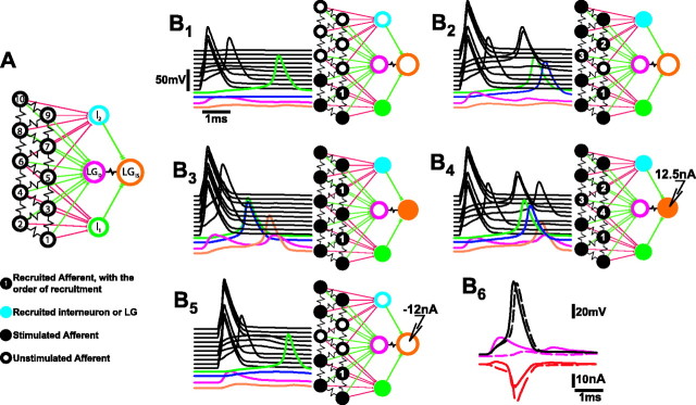
Fig. 8.
Electrical circuit model of the lateral excitatory network and LG. A, Each model compartment was characterized by reversal potentials and maximal conductances for sodium, potassium, and leakage currents. In all the compartments except LGD, which was passive, the sodium and potassium conductances were voltage- and time-dependent, as described by Hodgkin and Huxley (1952). B, Responses of the network to patterns of afferent stimulation. The 5 msec time course of each compartmental response is at left in each panel; interneuron and LG compartment responses are shown color-coded at thebottom; afferent responses are black and are arranged in ascending order according to the diagram inA. The pattern of stimulation and the network response are shown in the diagram at right in each panel, where brighter colored compartments are those that produced a spike.B1, Responses to simultaneous stimulation of compartments 1, 2, and 4 with 0.3 msec pulses of 300 nA depolarizing current. B2, Responses to stimulation of afferent compartments 1, 2, 4, 8, 9, 10.B3, Stimulation of all afferents except 3 and 7. B4, Afferents were stimulated according to the pattern used inB2 10 msec after onset of 12.5 nA current injection into LGIS.B5, Afferents were stimulated according to the pattern of B2 10 msec after onset of −12 nA current injection into LGIS.B6, The effect of the LGDEPSP on afferent recruitment. Afferents were stimulated according to the pattern of B2 when the network was intact (continuous traces) and when all afferent synapses to LGD except that of afferent 5 were removed (dashed traces). Top, Voltage responses (in millivolts) of afferent 5 and LGD.Bottom, Transynaptic current (in nanoamperes) through the rectifying electrical synapse between afferent 5 and LGD. Positive current moves from LGD to afferent 5.

