
Fig. 3.
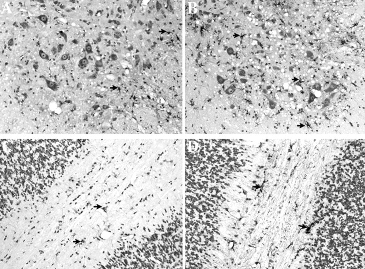
GFAP immunoreactivity is increased in G93A SOD1 mice lacking MT-I and MT-II. A, B, L5–L6 spinal cord ventral horn from 5.25-month-old G93A SOD1 transgenic mice with normal MT-I and MT-II (A) or absent MT-I and MT-II (B). C, D, Cerebellar white matter from 5.25-month-old G93A SOD1 transgenic mice with normal MT-I and MT-II (C) or absent MT-I and MT-II (D). Magnification, 200×.Arrowheads, GFAP reactive cells.