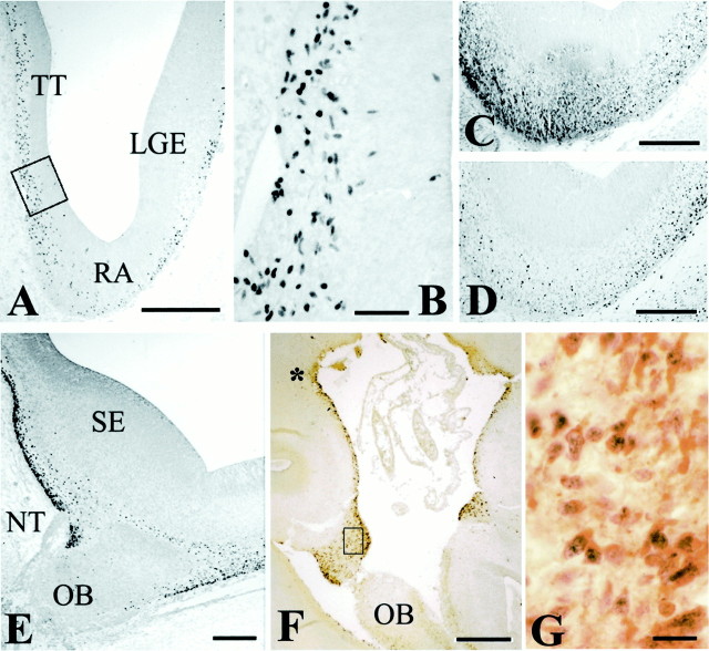
Fig. 3.
p73 and Reln in the periolfactory forebrain.A, CS 20 (6.5 GW). Panoramic view of the retrobulbar telencephalon, with p73-IR cells in taenia tecta (TT), retrobulbar area (RA), and lateral ganglionic eminence (LGE). B, High-power view of the boxed area in A, showing p73-IR cells in the VZ of TT. C, Reln;D, p73, CS 20. Parallel sections through the retrobulbar area showing partial dissociation of Reln and p73 expression.E, CS 22 (8 GW), p73. The medial periolfactory forebrain contains numerous p73-IR cells close to the septal eminence (SE). NT, Entrance of the nervus terminalis; OB, olfactory bulb. F, 14 GW. Panoramic view of the medial periolfactory forebrain, double staining p73/Reln. The asterisk marks the site where deep small p73-positive cells seem to ascend to the surface and begin to coexpress p73 and Reln. Mitral cells of the OB are Reln-positive but p73-negative. G, The boxed area inF. In the cell band surrounding the olfactory stalk, most cells coexpress p73 and Reln. Scale bars: A, 400 μm; B, 50 μm; C–E, 200 μm;F, 300 μm; G, 20 μm.

