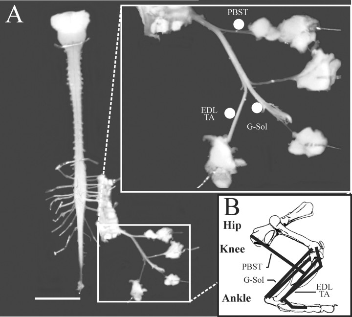
Fig. 1.
In vitro brainstem–spinal cord–sciatic nerve preparation. A, Ventral view of the preparation used in this study. The inset shows a higher magnification of the different nerve branches left attached to the spinal cord that are stimulated to identify motoneurons functionally.Filled circles indicate the approximate location of stimulating electrodes. Branches were identified by their muscular target (B). B, Schematic lateral view of the rat hindlimb showing the location and insertions of different muscles. EDL, Extensor digitorum longus;G–Sol, gastrocnemius–soleus;PBST, posterior biceps, semitendinosus;TA, tibialis anterior. Scale bar, 1 cm.

