
Fig. 9.
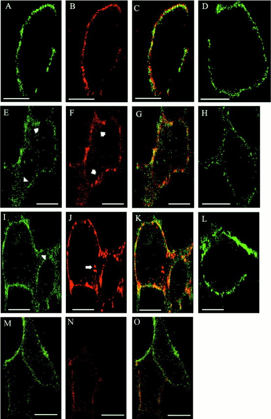
Antibody-induced endocytosis of L1 and β1 integrins in L1-expressing HEK cells. HEK293 cells were transfected for transient expression with L1(+RSLE) (A–C,E–G,I–K) or mutant L1(KGE) (M–O) plasmids. Confocal microscopy images of cells are labeled green for β1 integrin (A, E, I,M), red for wild-type or mutant L1 (B, F, G), andyellow for colocalized β1 integrin and L1 (C, G, K,O). Nontransfected cells were labeledgreen for β1 integrin alone (D,H, L). Live HEK cells were incubated with antibodies against L1 and β1 integrin for 0 min (A–D), 5 min (E–H,M–O), or 60 min (I–L). After 5 min, increased internal staining of β1 integrins was seen in L1-transfected cells (E,arrowheads) compared with nontransfected cells (H). Long arrow inJ indicated larger endocytotic vesicles of wild-type L1 resembling multivesicular bodies. L1(KGE) mutant cells (M) did not show increased β1 integrin internalization after 5 min. Images show representative cells from multiple experiments. Scale bars, 10 μm.