
Fig. 11.
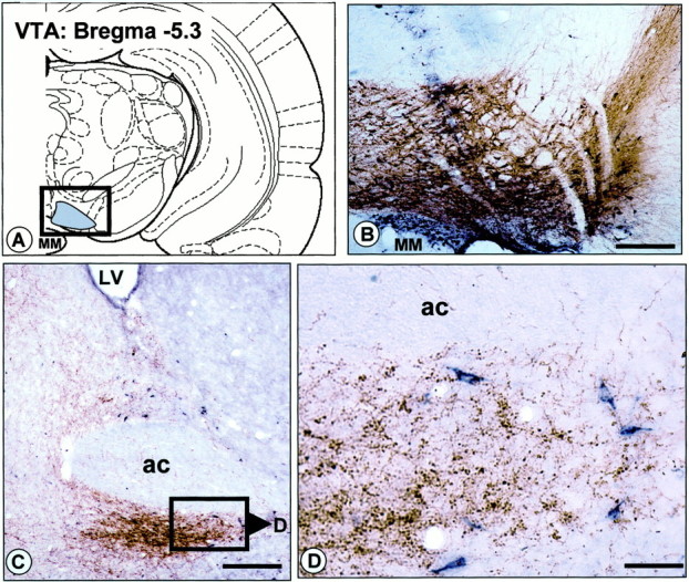
Projection from the vBNST to the VTA revealed by retrograde labeling in the vBNST after injection of CTb into the VTA.A, Schematic diagram illustrating the location of CTb injections in the VTA. B, Photomicrograph illustrating a representative CTb injection site in the VTA (dark bluelabeling). The section has been counterstained with TH immunohistochemistry (in brown), to delineate dopamine neurons and processes in the VTA (boxed area inA). C, D, Bright-field photomicrographs illustrating retrograde labeling in the vBNST after CTb injection into the VTA. The sections have been processed dually for CTb (dark blue) and DBH (brown). Note that numerous CTb+ cell bodies are observed in the dorsal and ventral BNST. Cell bodies retrogradely labeled in the vBNST are shown at higher power in D. ac, Anterior commissure; MM, medial mamillary nucleus. Scale bars:B, C, 1.0 mm; D, 0.1 mm.