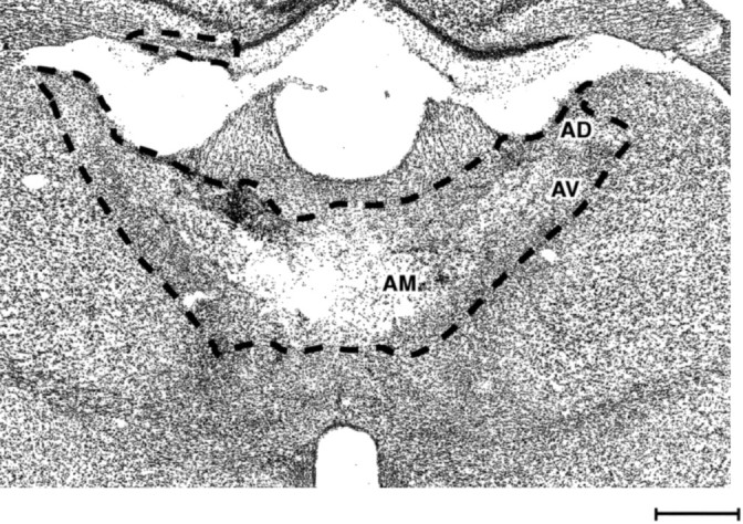
Fig. 3.

Photomicrograph of a Nissl-stained coronal section showing a bilateral thalamic lesion. The dashed line shows the extent of the lesion. The three principal anterior thalamic nuclei were the sole common lesion site in all cases. Scale bar, 500 μm.
