
Fig. 2.
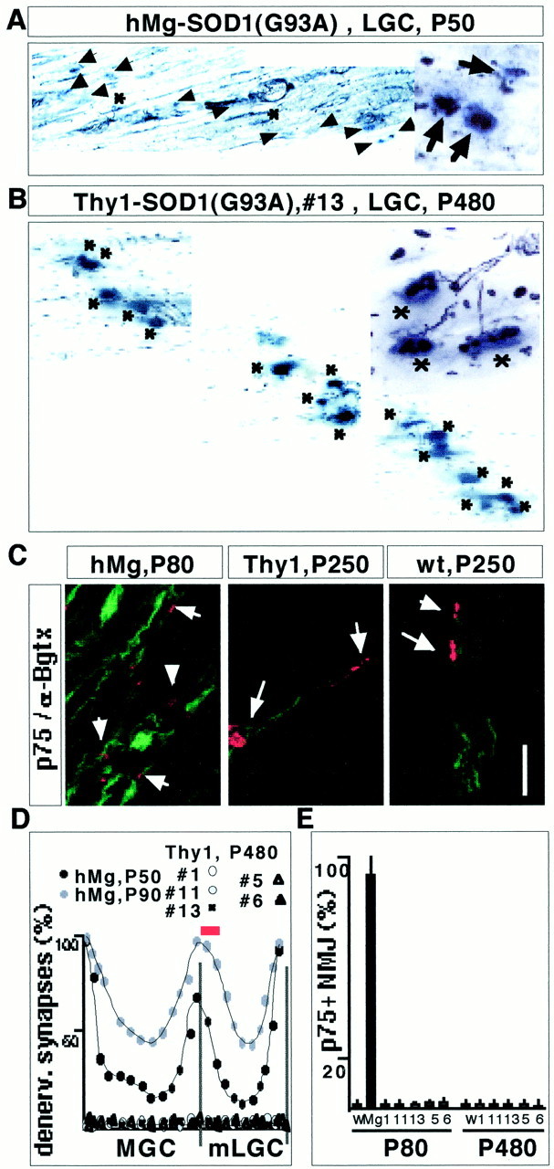
Absence of muscle denervation in Thy1-SOD1(G93A) and Thy1-SOD1(G85R) transgenic mice. A, B, Combined silver esterase reaction of LGC [medial end of medial compartment (mLGC), as indicated by the red bar inD] from an hMg-SOD1(G93A) mouse at P50 (A) and a Thy1-SOD1(G93A) line 13 mouse at P480.Insets on the right show a detail from the same region. Synaptic regions are in blue(acetylcholine esterase reaction product), and nerves are inblack. Some of the nuclei on the section yield ablack background color. Asterisks, Innervated NMJs; arrows, denervated NMJs.C, Double-labeling immunocytochemistry for p75 and α-bungarotoxin in LGC (medial compartment). The Thy1 transgenic mouse was from the Thy1-SOD1(G93A) 13 line. Arrows point to some of the α-bungarotoxin-positive NMJs. The weak p75 signal on the sections from wild-type and Thy1 transgenic mice was associated with sensory nerves, whereas the strong signal in the hMg mice was attributable to Schwann cells that had lost contact with motor nerve. The small size of the acetylcholine receptor cluster signals in the hMg mouse is attributable to chronic denervation of mLGC in this mouse. Scale bars: A, B, 120 μm; insetin A and B, 50 μm; C, 60 μm. D, Quantitative analysis of denervation (denerv.) along the synaptic band of MGC and LGC muscle (see Materials and Methods for details). Each curve represents average values from two mice. E, Quantitative analysis of mLGC denervation (p75-positive NMJs; see Materials and Methods for details). W, Wild type; Mg, minigene, hMg-SOD1(G93A). Average values from two mice each are shown.