
Fig. 3.
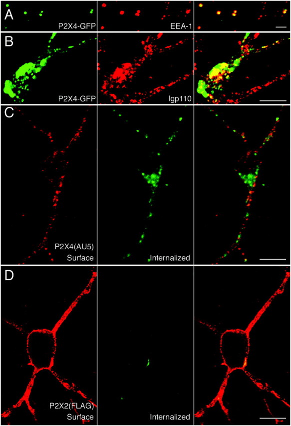
P2X4, but not P2X2, receptors are constitutively internalized. In distal processes, P2X4–GFP puncta colocalize with anti-EEA1/Cy3 staining (A), and costaining with anti-lgp110/Cy3 shows that large accumulations of P2X4–GFP in the cell soma are within lysosomes (B). Live-labeling of P2X4(AU5) (C) and P2X2(FLAG) (D) receptors for 30 min at 37°C with anti-AU5 and anti-FLAG, respectively. In C and D, cell surface (left) and internalized (middle) receptors were visualized using Cy3-conjugated and FITC-conjugated secondary antibodies, before and after permeabilization, respectively. Overlaid images are shown on the right panels. Scale bars: A, 2 μm;B–D, 10 μm.