Fig. 4.
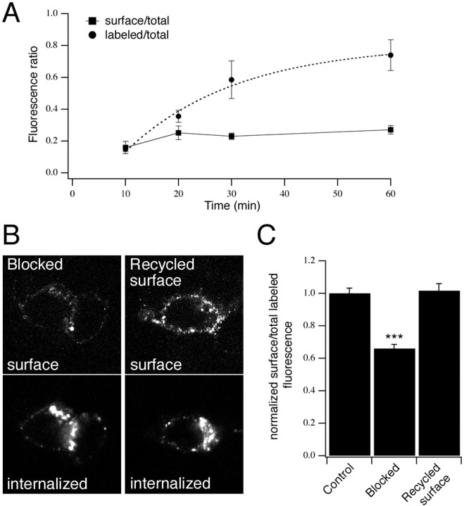
Cycling of P2X4(AU5) receptors to and from the surface. A, Time course of P2X4(AU5) constitutive internalization in neurons. Transfected neurons were labeled with anti-AU5 for different lengths of time at 37°C. Cells were stained using secondary antibody before (surface/total) or after (labeled/total) permeabilization and then stained for total P2X4 receptor. Fluorescence ratios are shown plotted against antibody incubation time. The time course of labeling is described by a single exponential (τ = 21 min;n = 4–7 neurons for each time point).B, C, Internalized P2X4(AU5) receptors recycle back to the plasma membrane. Representative confocal images used for analysis show an increase in P2X4(AU5) receptors recycled to the cell surface (B). Histogram (C) shows the surface fluorescence as a fraction of total (surface plus internalized) fluorescence, normalized toControl cells that were fixed immediately after a 30 min incubation with anti-AU5. Blocked represents cells that were fixed immediately after incubating cells with a nonfluorescent secondary antibody at 4°C. Recycled surface represents cells that were returned to 37°C for 15 min after blocking. Surface and internalized receptors were detected before and after permeabilization using Cy3- and FITC-conjugated secondary antibodies, respectively (n = 14–22 neurons for each condition).

