
Fig. 6.
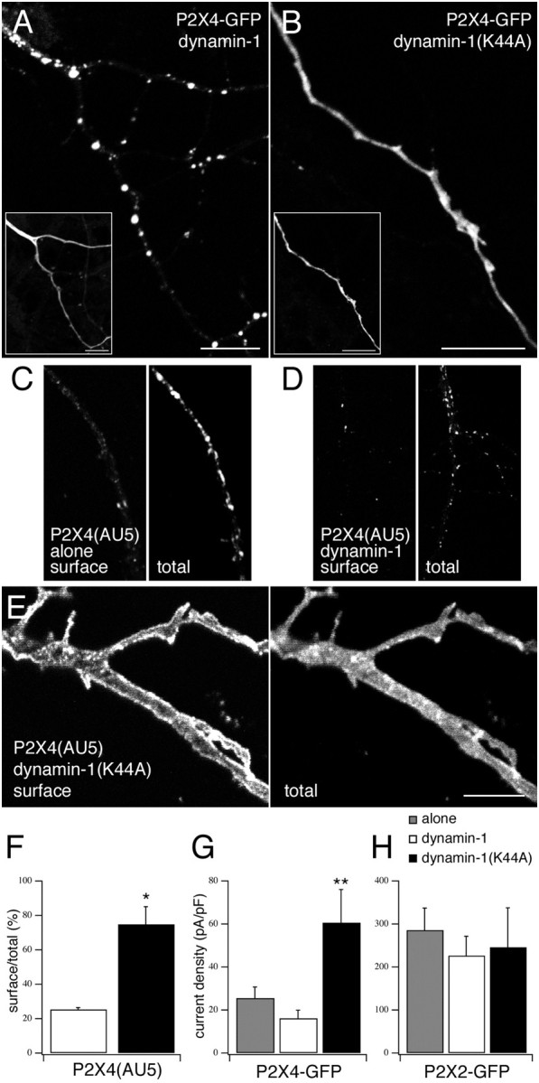
Trafficking of P2X4 is dynamin-dependent.A, B, Confocal images of P2X4-GFP coexpressed with either wild-type (A) or dominant-negative (B) dynamin-1.Inset images show detection of the coexpressed dynamin-1 constructs using anti-HA/Cy3. C–E, Confocal images to show neurons expressing P2X4(AU5) alone (C) or with either wild-type dynamin-1 (D) or dynamin-1(K44A) (E) were immunostained for cell surface receptors (anti-AU5/Cy3 before permeabilization) and total receptor (anti-P2X4/FITC after permeabilization). Scale bars, 10 μm.F, Quantification of surface versus total receptor in neurons coexpressing P2X4(AU5) and either dynamin-1 or dynamin-1(K44A) (n = 3 neurons for both condition).G, H, Histograms of the mean whole-cell current densities after application of ATP (100 μm) in neurons expressing either P2X4–GFP (G) or P2X2–GFP (H) alone (shaded) or coexpressed with either dynamin-1 (white bars) or dynamin-1(K44A) (black bars) (n = 5–15 neurons for each condition).