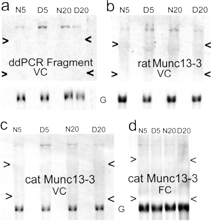
Fig. 3.
Differential expression of Munc13-3. Northern blots confirming differential expression of the candidate gene Munc13-3. Total RNA from a 5 week normal (N5), a 5 week dark-reared (D5), a 20 week normal (N20), and a 20 week dark-reared (D20) cat visual cortex (a–c) or frontal cortex (d) was loaded in each blot. Blots were either rehybridized (a, b) with GAPDH (G) or simultaneously hybridized (c, d) with a Munc13-3 probe and GAPDH.a, Filter of visual cortical (VC) total RNA hybridized with the 3′ cat Munc13-3 gene fragment recovered from ddPCR; b, filter of VC total RNA hybridized with a full-length rat Munc13-3 probe; c, filter of VC total RNA hybridized with a probe designed for the 5′ end of the cat Munc13-3 gene (see Materials and Methods); d, filter of frontal cortical (FC) total RNA hybridized with the same 5′ cat Munc13-3 probe. All probes identified a band of appropriate size (∼7 kb) for Munc13-3, based on the rat sequence. Arrowheadsindicate 28S and 18S rRNA.

