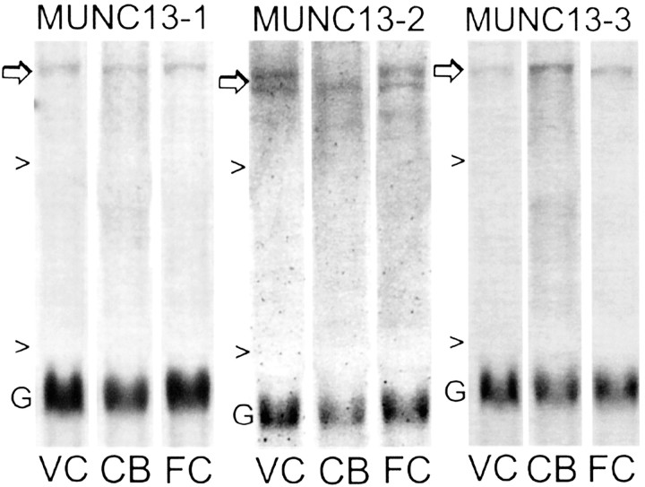
Fig. 4.
Differential regional distribution of Munc13 mRNAs. Portions of Northern blots showing regional differences in expression of Munc13 family members (Munc13-1, Munc13-2, Munc13-3) in two neocortical structures [visual cortex (VC) and frontal cortex (FC)] and cerebellum (CB) of normal 20 week cats. Arrows indicate the Munc13 band of interest, and arrowheads indicate 28S and 18S rRNA. Blots were simultaneously hybridized with the Munc13 probe and with the probe to GAPDH (G). Probes were 5′ cDNAs specific for each Munc family member, and they share 98–99% sequence identity between rats and cats (see Materials and Methods).

