
Fig. 2.
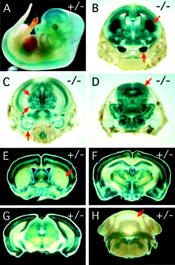
Pattern of fz3 expression as determined by X-gal staining of the knocked-in lacZreporter in fz3(+/−) and fz3(−/−)mice. A, fz3(+/−) embryos at E12 show widespread X-gal staining in the developing CNS. B–D,fz3(−/−) embryos at E18; coronal series from rostral (B) to caudal (D).B, Centered on the striatum; C, centered on the thalamus; D, centered on the tectum. Widespread X-gal staining is seen in the cerebral cortex, diencephalon, and brainstem, with the highest levels in the developing striatum (arrow at 45° angle in B) and the trigeminal ganglia (vertical arrow in B). Staining is also seen in the inner ear (vertical arrowin C), dorsal and ventral thalamic nuclei (45°arrows in C), tectum (arrow in D), and retina (data not shown). fz3(+/−) embryos at E18 show a nearly identical pattern of X-gal staining but at lower intensity. E–H, Adult fz3(+/−) brains show X-gal staining in the cerebral cortex and select midbrain structures with minimal staining in the striatum (arrow in E), cerebellum (arrow in H), and brainstem. Coronal series from rostral (E) to caudal (H). E, Centered on the striatum; F, centered on the thalamus; G, centered on the colliculus; H, centered on the cerebellum.