
Fig. 6.
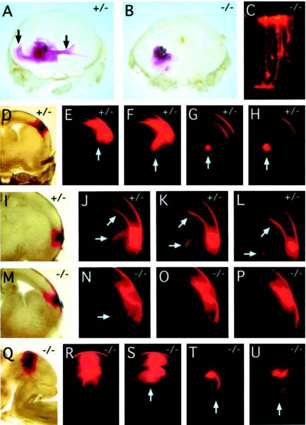
DiI labeling of cortical, commissural, and tectal fiber tracts at E18. A, DiI crystals placed in the caudal region of the striatum of a fz3(+/−) brain label the external capsule and the genu of the corpus callosum in the ipsilateral cortex and the anterior commissure in the contralateral hemisphere (vertical arrows). B, In thefz3(−/−) brain, there is little diffusion of the DiI beyond the site of crystal placement. C, Individualfz3(−/−) cortical neurons labeled with microcrystals of DiI from the cortical surface (top) show normal morphologies. D–L, Serial coronal sections through twofz3(+/−) brains in which DiI crystals were placed in the dorsolateral (D) or lateral (I) cortex. Prominent labeling is seen in cortical fibers traversing the internal capsule (vertical arrows in E–H and horizontal arrows in J–L) and in the genu of the corpus callosum (arrows at 45° angle in J–L).M–P, Serial coronal sections through afz3(−/−) brain in which DiI crystals were placed in the lateral cortex. The DiI has spread locally but has labeled only a small number of fibers traversing the internal capsule (arrow in N). Q–U, Serial sagittal sections through an fz3(−/−) brainstem in which DiI crystals were placed in the lateral tectum.R–U, Efficient labeling of the tectospinal tract (arrows in S–U) with midline crossing in T, a labeling pattern indistinguishable from that seen in fz3(+/+) or fz3(+/−)brains. Sections are 300 μm in thickness.