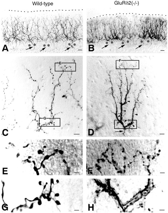
Fig. 2.

Anterogradely labeled CFs in lobule VI of the wild-type (A, C, E, G) and GluRδ2 knock-out (B, D, F, H) cerebella. A, B, Low-power views of cerebellar regions with massive CF labeling. The pial surface is indicated by the dotted line.Arrows indicate thin labeled axons, which run through the granular layer and between PC somata (asterisk).C, D, CFs with isolated labeling.Arrowheads in D indicate the dense, plexus-like innervation characteristic of the knock-out mouse.E, F, High-power views of the top portions in C and D. CF tendrils having small terminal boutons are increased in the knock-out mouse.G, H, High-power views of the bottom portions in C and D. A dense, plexus-like innervation is conspicuous in the knock-out mouse. Scale bars: A, B, 20 μm; C, D, 10 μm;E–H, 2 μm.
