
Fig. 5.
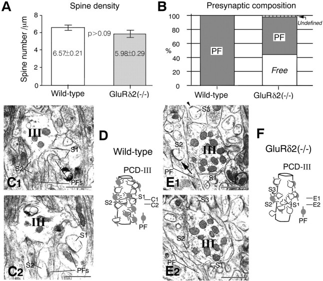
PCD-III (distal) domain. A, Spine density. B, Presynaptic composition on PC spines.C1, C2, Serial electron micrographs in the wild-type mouse. All spines, including S1 and S2, are contacted by PFs. E1, E2, Serial electron micrographs in the knock-out mouse. Spine S2 is contacted by PF, but spines S1 and S3 are free of innervation. The arrow in E1indicates elongated postsynaptic density that exceeds over the synaptic junction between the PF terminal and spine S2. D, F, Reconstructed images of a part of the PCD-III domain. Scale bars, 1 μm.