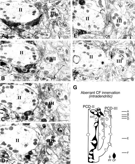
Fig. 6.

Serial electron micrographs showing aberrant CF innervation against adjacent spiny branchlets of the same PC in the GluRδ2 knock-out mouse. A labeled CF ascending the PCD-II dendrite (II) forms synaptic contact with spineS2 (B, C). Spine S2protrudes from an adjacent spiny branchlet (D, III), which is branched from that PCD-II dendrite in a deeper region of the molecular layer (F, arrow). SpinesS1 and S3 protrude from the PCD-III or PCD-II dendrite and are innervated by PF or labeled CF, respectively.Lines emitting from marked spines point to either a spine head or spine neck connecting to dendrites. G, Reconstructed image of the “intradendritic type” of aberrant CF innervation. Scale bars, 1 μm.
