
Fig. 3.
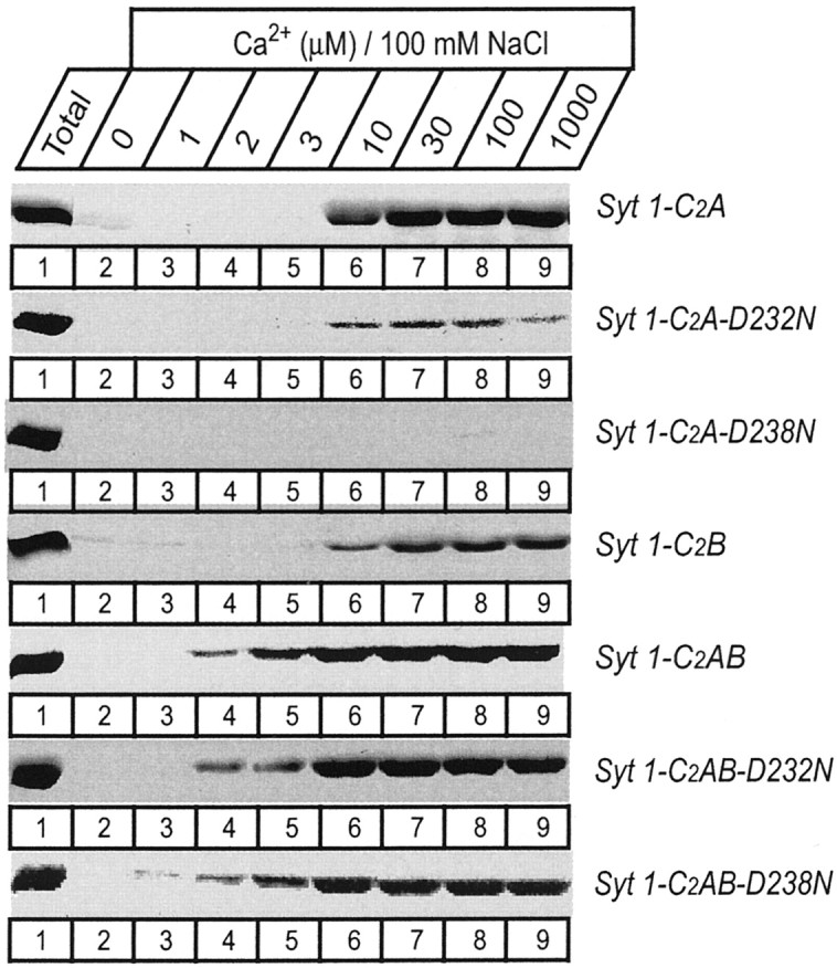
Apparent Ca2+ affinities of wild-type and mutant single and double C2 domain fragments from synaptotagmin 1 measured by Ca2+-dependent binding to liposomes. The isolated wild-type and D232N and D238N mutant C2A domain, the wild-type C2B domain, and the wild-type and mutant double C2 domain fragments were analyzed. Liposomes composed of 25% PS/75% PC were incubated with the indicated C2 domains (present as soluble purified GST-fusion proteins) at the Ca2+ concentrations shown on top (clamped with Ca2+/EGTA buffers) and centrifuged, and bound proteins were analyzed by SDS-PAGE and Coomassie blue staining. Data shown are from a single representative experiment repeated multiple times.