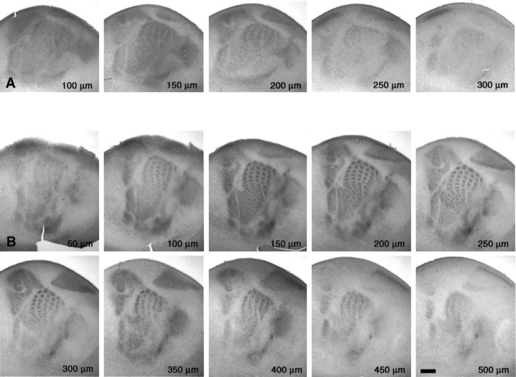
Fig. 3.
Emergence of periphery-related patterns is observed most clearly in the upper layers of the cerebral cortex, as viewed from serial tangential sections through S1. The hemispheres were flattened between two glass slides and sectioned in the tangential plane to 50-μm-thick sections. The serial order was maintained throughout the 5-HTT immunohistochemical procedure. The distance from the pial surface was estimated by counting the number of sections from the first section through the pia matter. Two sets are shown at P3 and P5. TCA patterning is most clearly visible at 150 μm below the pial surface at P3 and at 200 μm below the pial surface at P5, which corresponds to the position of layer IV at that age. Scale bar, 500 μm.

