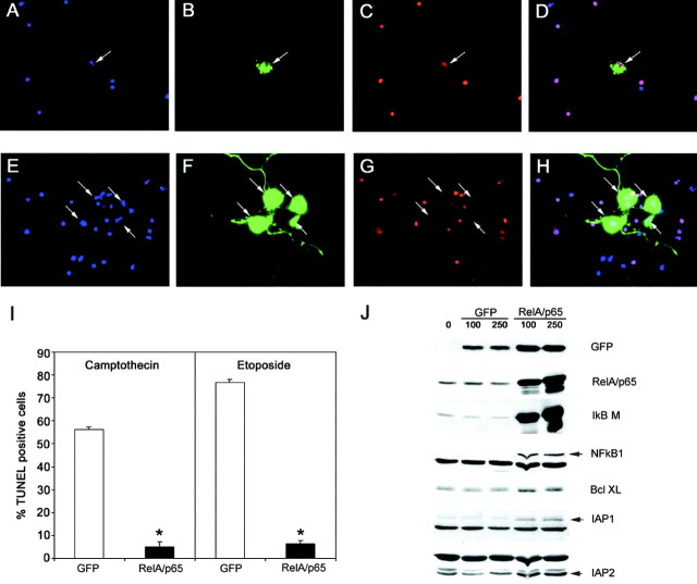
Fig. 6.
p65/RelA protects cortical neurons from apoptotic death. E15–16 cortical neurons were infected with 75 MOI of recombinant adenovirus encoding GFP alone (A–D) or with recombinant virus encoding both p65/RelA and GFP (E–H) for 24 hr. Cells were then exposed to etoposide (20 μm) for an additional 18 hr and then fixed and analyzed for GFP fluorescence (B, F,green), for apoptosis using Hoescht 33342 nuclear staining (A, E, blue), and TUNEL labeling (C, G,red). D, H, Merged images of A–C and E–G, respectively. Cells infected with GFP alone (and uninfected cells) rapidly underwent apoptosis when exposed to etoposide, whereas neurons infected with p65/RelA were robustly viable under these conditions. I, Cells were infected with 75 MOI of adenovirus expressing either GFP or expressing GFP together with p65/RelA for 48 hr and then treated with camptothecin (20 μm) or etoposide (20 μm) for 18 hr. GFP-positive cells were scored for TUNEL-positive nuclei. Expression of p65/RelA conferred robust protection from apoptosis because of campthothecin (*p < 0.001) or etoposide (*p < 0.0001). At least 300 cells were assessed for each condition, and results were analyzed for statistical significance by Student's t test. J, E16 cortical neurons were either left uninfected or were infected with 75 MOI of recombinant adenovirus expressing GFP or expressing both GFP and p65/RelA for 48 hr. Neurons were then lysed and analyzed by immunoblot. Levels of endogenous IκBα, NFkB1, IAP1, IAP2, and Bcl-XL were specifically increased by p65/RelA overexpression.

