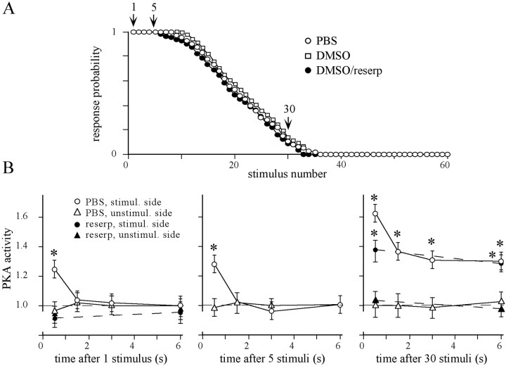
Fig. 2.
Habituation of the proboscis extension response and the dynamic of PKA activation in the antennal lobes. A, Animals were injected with 1 μl of PBS, DMSO, and DMSO containing 2 μg of reserpine 18 hr before habituation. Sucrose stimuli were applied to one antenna until the habituation criterion was fulfilled. The data points for each stimulus represent the average of the response probability of all animals in the respective group (20 animals each). Habituation kinetics did not differ between the groups (ANOVA, p = 0.95). B, PKA activity in the antennal lobes of the stimulated and the unstimulated sides was determined at different times after the 1st, 5th, and 30th stimulus (see arrows inA). In each experiment, values were normalized to PKA activity in unstimulated animals, which are defined as the relative PKA activity of 1 (dashed lines). Each data point represents the mean ± SEM of relative PKA activity of at least eight independent measurements [1st and 5th stimulus: ANOVA, p < 0.0001; 0.5 sec after stimulation, the PBS-injected group significantly differs from the corresponding unstimulated AL (*p < 0.001, pairedt test) and all other means (p < 0.003, unpaired ttest); 30th stimulus: ANOVA, p < 0.0001; at each time point, the means of the stimulated ALs (PBS- and reserpine-injected groups) significantly differ from their respective unstimulated ALs (*p < 0.001, pairedt test)]. Value at 0.5 sec (PBS-injected animals) significantly differs from all other values (p < 0.003, unpaired ttest).

