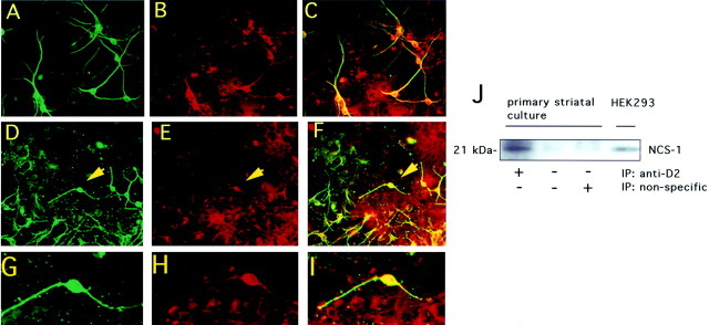
Fig. 3.
Expression of NCS-1 and D2 receptors in primary striatal neurons from rat brain. Detection of anti-MAP2 (A) and anti-NCS-1 (B) in double labeling within neurons. Merged image (C) shows MAP2 and NCS-1 coexpression within neuronal perikarya and dendritic process of virtually all striatal neurons. Detection of D2 receptors (D) and NCS-1 (E) in double-labeled cultures. Merged image (F) shows D2 receptor and NCS-1 coexpression within the cell body and processes of neurons. High magnification of a representative neuron (indicated by arrowheads in D–F) showing expression of D2 receptors (G), NCS-1 (H), and colocalization of the two proteins (I). Numerous D2-positive, NCS-1-negative cells display an apparent astrocytic morphology (D–F). J, Interaction of D2 receptors and NCS-1 in striatal cultures Anti-D2 receptor antibody was used to immunoprecipitate D2 receptors from crude membrane preparations of striatal cells. Immunocomplexes were then probed with anti-NCS-1 antibodies. The position of NCS-1 endogenously expressed in HEK 293 cell membranes is shown (HEK 293 lane).

