
Fig. 4.
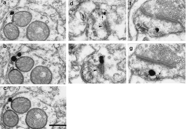
NCS-1 and D2 receptors colocalize within dendritic shafts and spines. Representative images of immunolabeling of NCS-1 (DAB reaction product) and D2 (immunometal particles) receptors within dendritic shafts (a–c) and spines (d–g)of neurons within monkey (a–e) and rat (f, g) striatum. Silver grains (representing D2 receptors) were detected extrasynaptically (a–d, f, g)and perisynaptically (e). Within dendritic spines, the spine apparatus appears to be immunoreactive for NCS-1 (d–g). D2 receptor immunoreactivity is associated with the membrane of the NCS-1-immunoreactive spine apparatus (f, g). Note the close spatial contiguity of DAB deposits (arrows) and silver particles in the dendrite as well as within spines (b, d, g). An immunonegative spine is present in the top right corner ofa–c. Arrowheads point to the postsynaptic density of asymmetric synaptic contacts (d, e). Scale bar, 200 nm.