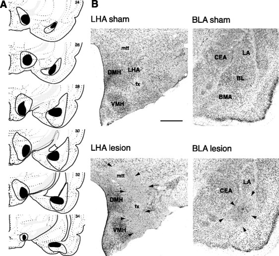
Fig. 1.

Histology. A, The extent of the largest (enclosed black area) and smallest (filled black area) acceptable lesions at several rostrocaudal levels for all rats in the contralateral and ipsilateral groups. Except for minor mechanical damage along the injector tracks, no damage was evident in any of the sham-lesioned brains. Plates adapted from the atlas of Swanson (1992). B, Representative photomicrographs of lesion and sham histology.Arrows denote lesion borders. Scale bar, 0.5 mm. Amygdala: BLA, Basolateral area (includesBL, BMA, and LA);BL, basolateral (basal); BMA, basomedial (accessory basal); CEA, central; LA, lateral nuclei. Hypothalamus: DMH, Dorsomedial nucleus;fx, fornix; LHA, lateral area;mtt, mammillothalamic tract; VMH, ventromedial nucleus.
