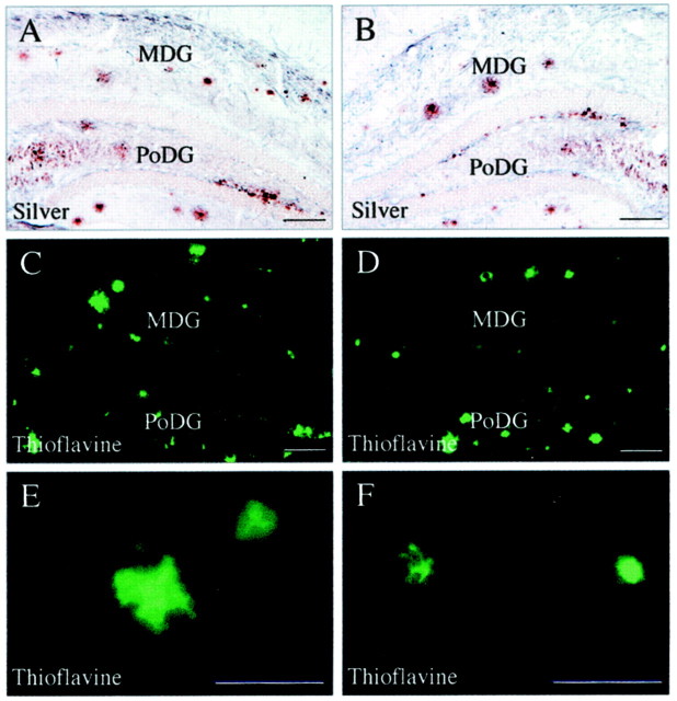
Fig. 5.
Effects of ERC lesions on amyloid plaques visualized with traditional histochemical methods such as silver (A, B) and thioflavine (C–F). Lesioned side is on theright. There is a trend for a reductions of both silver- and thioflavine-stained plaques in the molecular layer of the dentate gyrus. There are no obvious differences in the polymorph cell layer.MDG, Molecular layer of dentate gyrus;PoDG, polymorph layer of dentate gyrus. Scale bars:A–D, 100 μm; E, F, 50 μm.

