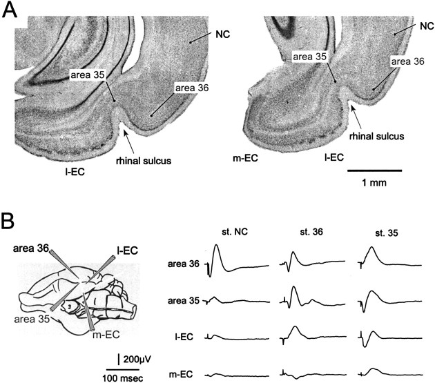
Fig. 1.
A, Coronal sections of the rhinal region of the guinea pig at two different rostrocaudal levels. The NC, area 36, and area 35 in the perirhinal cortex, the m-EC, and the l-EC are indicated. The rhinal sulcus is indicated by thearrowhead. B, Field responses evoked in area 36, area 35, l-EC, and m-EC by surface stimulation of the NC (left column), area 36 (middle column), and area 35 (right column). A schematic drawing of the positions of the recording electrodes is shown on theleft. All recordings were performed at 500–600 μm depth.

