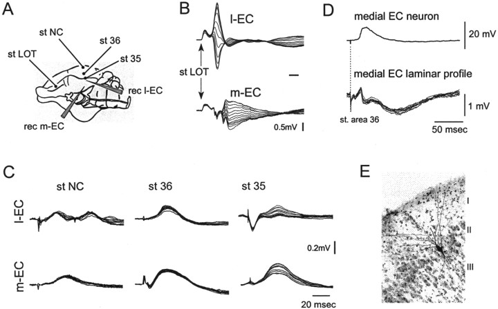
Fig. 4.
Field potential depth profiles recorded in m-EC and l-EC in response to stimulation of superficial NC, area 36, and area 35. A scheme of the position of the electrodes is illustrated inA. As illustrated in B, to guide the placement of the recording probes, we used the responses to LOT stimulation, characterized by potentials with different delays that showed a reversal within the cortical profile (Biella and de Curtis, 2000) in m-EC and l-EC. Note that reversed potentials in l-EC have a shorter delay than in m-EC. C, Superimposed field responses to stimulation in NC, area 36, and area 35 recorded simultaneously with the 16 leads of a silicon probe inserted in the l-EC (top traces) and the m-EC (bottom traces). D, Simultaneous intracellular and field potential profile recorded in the medial EC after stimulation of superficial layer in area 36. The intracellularly recorded neuron was filled with byocitin at the end of the electrophysiological recording and was identified as a layer II stellate neuron of the medial EC on the thionin-counterstained 79-μm-thick section illustrated inE.

