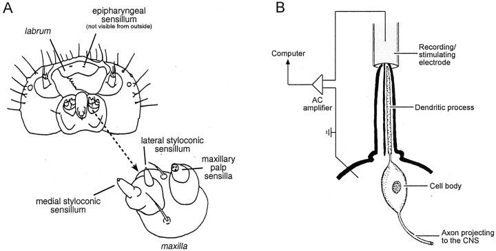
Fig. 1.
A, Diagram of the head of M. sexta, as viewed from below. An enlargement of a maxilla (indicated with an arrow) is provided to show the location of the medial and lateral styloconic sensilla. The epipharyngeal sensilla are located underneath the labrum and thus are not visible in this diagram. There are four taste cells in each of the lateral and medial styloconic sensilla, three taste cells in each of the epipharyngeal sensilla, and three to four taste cells in each of the gustatory sensilla on the maxillary palp. One of the taste cells in each sensillum is bitter sensitive. [This illustration was adapted from Bernays and Chapman (1994), their Fig. 3.4.] B, Diagram of the tip recording method (Hodgson et al., 1955) for recording excitatory responses of taste cells (or neurons) located within a single taste sensillum (for examples, see Fig.2A). During a recording, the tip of a taste sensillum is inserted into the end of a glass recording/stimulating electrode, which is filled with an electrolyte solution (0.1m KCl in deionized water) and the taste stimulus. The stimulus solution diffuses through a pore in the tip of the sensillum and activates a transduction mechanism(s) on the distal end of the dendritic process of the taste cell; the electrode records the ensuing action potentials. For clarity, only one taste cell is indicated. Note that the axonal process of the taste cell projects directly to the CNS.

