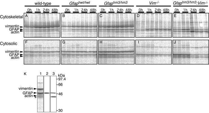
Fig. 6.
Pulse-chase experiments of GFAP. Primary cultured astrocytes prepared from wild-type (A,F), Gfaphwt/hwt(B, G),Gfaphm3/hm3 (C,H), Vim−/−(D, I), andGfaphm3/hm3:Vim−/−(E, J) embryos were labeled for 15 min with 35S-Met/Cys, followed by immediate harvesting or chasing for 1, 24, or 48 hr. Autoradiographs of Triton X-100-insoluble cytoskeletal fractions and immunoprecipitates from Triton X-100-soluble cytosolic fractions with anti-GFAP antibody are shown inA–E and F–J. K, Lower molecular weight product detected by the GFAP antibody (asterisk) was not detected using a head domain-specific antibody. Immunoprecipitates fromGfaphm3/hm3 astrocytes were analyzed using autoradiography (lane 1), immunoblot with antibody to GFAP head domain (lane 2), or immunoblot with GF 12.24 as pan-GFAP antibody (lane 3).

