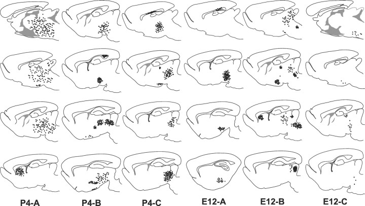
Fig. 1.
Distribution of postnatal (P4-A–C) and embryonic (E12-A–C) cerebellar cells transplanted to the embryonic rat brain in utero. For each case, four serial sections, modified from Paxinos and Watson (1982), are shown (top to bottom = medial to lateral). Each dotrepresents cellular aggregates or groups of single scattered cells.

