
Fig. 3.
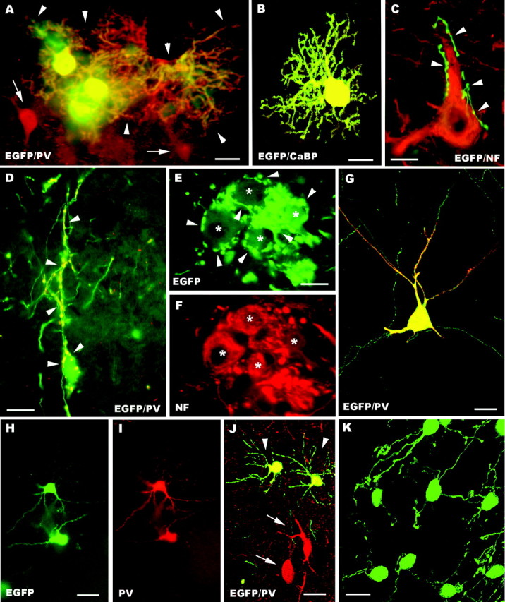
A–K, Fate of embryonic cerebellar cells engrafted in ectopic regions of the embryonic brain in utero. Parvalbumin-immunolabeled Purkinje cells with large dendritic trees (arrowheads) are illustrated inA; arrows point to adjacent parvalbumin-positive host neurons. Another calbindin-immunolabeled transplanted Purkinje cell, bearing less extended dendrites, is shown in the confocal image B. In C a presumptive EGFP-positive Purkinje axon (arrowheads), recognized by the particularly intense fluorescence, terminates on a neurofilament immunostained host neuron. Frequently, Purkinje axons (arrowheads in D point to the parvalbumin-immunolabeled terminal branches) enwrap the somatodendritic surface of EGFP-positive multipolar neurons. E and Fshow a small cluster of these neurons (asterisks), which are labeled by anti-neurofilament antibodies (F) and covered by strongly fluorescent Purkinje axons (arrowheads in E). The morphological features of EGFP-multipolar neurons, bearing slender dendrites with long ramifications, are illustrated in the confocal pictureG; note that this neuron is also double labeled for parvalbumin. H and I show small parvalbumin-immunopositive neurons, classified as molecular layer interneurons. Arrowheads in J point to another two of such neurons settled in the host striatum; note the different size and morphology shown by transplanted and recipient neurons (arrows). A cluster of transplanted granule cells in the host dorsal cochlear nucleus is displayed by the confocal picture K. NF, Neurofilament SMI32; PV, parvalbumin; CaBP, calbindin; EGFP, enhanced green fluorescent protein. Scale bars: K, 10 μm; C, E, F, 15 μm; B, D, 20 μm;A, 25 μm; G–J, 30 μm.