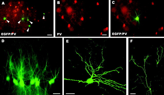
Fig. 5.
A–F, Fate of embryonic neocortical precursors grafted to the embryonic CNS in utero. Micrograph A shows EGFP-positive embryonic neocortical cells (some are pointed by arrowheads) engrafted in the host neocortex. Some of such neurons (B, C) show the morphology of stellate cells and can be immunolabeled by anti-parvalbumin antibodies. Many of these grafted cells show site-specific morphological features, such as presumptive pyramidal neurons in hippocampal CA1 (D), medium-sized spiny neurons in the striatum (E), and olfactory bulb interneurons (F). PV, Parvalbumin, EGFP, enhanced green fluorescent protein. Scale bars: B–E, 20 μm; A,F, 50 μm.

